Posts
-
How to build a Fiber Photometry System
At its core, fiber photometry (FIP) is a method used to record and measure changes in fluorescence signals within specific neuronal populations in the living brain. These fluorescence signals can be elicited by genetically encoded calcium indicators (GECIs) or other fluorophores, which glow brighter when a neuron fires. By channeling light through optical fibers directly to a target brain region, and then collecting and analyzing the emitted light in response, scientists can track neuronal activity in real-time.
FIP is minimally invasive and highly popular. However, off-the-shelf FIP systems can be costly and inflexible which is why open-source systems such as the one presented in this paper were developed. These systems have the advantage that they can record from multiple brain regions simultaneously and that they can be flexibly modified. In addition, building your own system has the important advantage that you’ll become intimately familiar with the mechanics of FIP.
In this walk-through we’ll go over how build a camera-based FIP system. I’ll also share code for the acquisition and analysis of FIP recordings.
Read more -
Visualizing COVID
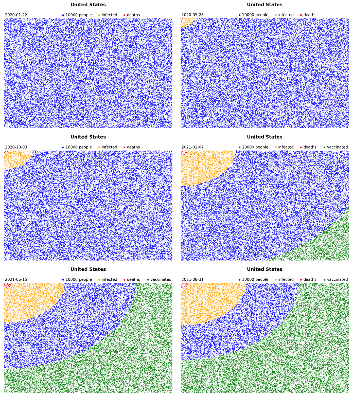
“2 million people died of COVID” or “The US has surpassed 25 million cases” - Despite the fact that more people die from COVID every day than died on 9/11, these numbers just don’t seem to make the impression anymore like they did at the beginning of the pandemic. I made these animations to put these numbers into perspective, and to try and wrap my head around them. If you’re interested in making your own, I share the code below.
These blue squares represent the entire population of a country. Every blue dot stands for a certain number of people. Every orange dot represents that same number of people after they are infected with COVID, while the red dots refer to the COVID death toll.
Read more
-
The Beauty of the Dopamine system
Ex-vivo Slice Electrophysiology (commonly known as ‘patching’) is a technique whereby we insert a tiny patch pipet into a neuron in order to record from it. It’s a very involved process that requires a lot of practice. Patching your first neuron is generally cause for cellebration. I’ve used slice electrophysiology to study the effects of cocaine administration on synaptic plasticity on projection-defined dopamine neurons. Since it was important to me to be able to identify which exact neuron I recorded from, I filled my pipets with a dye called biocytin. While recording from dopamine neurons the biocytin difuses into the cell, which allowed me to later inspect the cell underneath a microscope. The images below are all dopamine neurons obtained in this matter. They are in a brain area between the brain stem and the cerebrum called the Ventral Tegmental Area (VTA).
Read more


